
What Is Trichoscopy? Scalp Dermoscopy Explained
What is trichoscopy? A plain guide to scalp dermoscopy: how a trichoscope works, what it shows, who uses it, and how it compares with a biopsy or pull test.
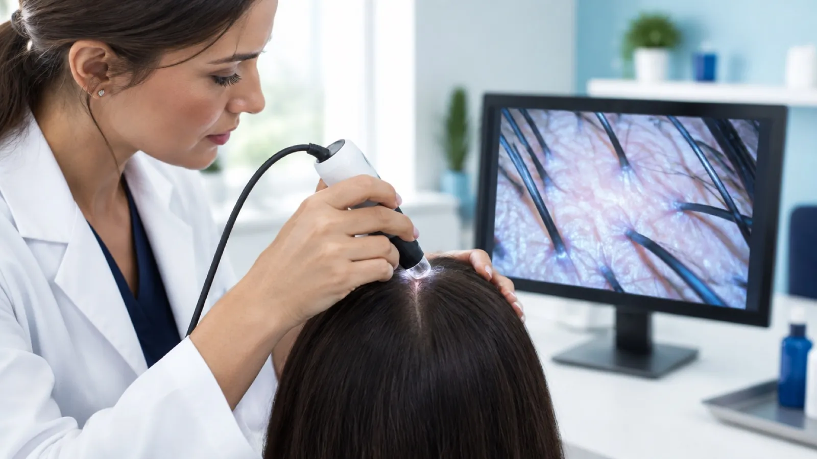
What is trichoscopy? Trichoscopy is the examination of the hair and scalp using a dermoscope, a lighted magnifier held on or near the skin. It is noninvasive, takes a few minutes, and shows follicular openings, hair shaft thickness, scale and blood vessels that the eye cannot see unaided.
Trichoscopy is one of the core observation skills in trichology. This guide covers the trichoscopy meaning, how the device works, what a trichoscope shows, who uses it and for what, how it compares with a scalp biopsy and a hair pull test, and how to choose a scope. USTI content is educational and non-medical.
What is trichoscopy?
DermNet defines trichoscopy as examination of the scalp and hair using a handheld or a videodermoscopy device. A review in Skin Appendage Disorders by Ocampo-Garza and Tosti puts it in one line: trichoscopy is "the dermoscopic examination of the hair and scalp."
A few related words:
- Trichoscope. The device. DermNet describes a handheld trichoscope as a conventional dermatoscope.
- Scalp dermoscopy. Another name for trichoscopy.
- Scalp scope. The everyday salon term for a digital magnifying camera used on the scalp.
The method is fairly recent. A 2008 paper in the Journal of Drugs in Dermatology by Rudnicka and colleagues was titled "Trichoscopy: a new method for diagnosing hair loss." For definitions of the terms used below, keep the trichology glossary open.
How does trichoscopy work?
Trichoscopy works by combining magnification with a controlled light source so the scalp surface and the hair shafts can be seen in detail. Three things vary between devices: the magnification, whether the device is handheld or video, and whether the light is polarised.
Magnification
DermNet lists typical handheld dermoscope magnifications of 10x, 16x or 40x, and gives a range of x20 to x160 for trichoscopy. A 2009 review of hair evaluation methods in the International Journal of Trichology notes that images are usually obtained at 20x to 70x.
Handheld vs video
| Handheld dermoscope | Video dermoscope or digital scalp scope | |
|---|---|---|
| How you view | Through the lens, or through a phone or camera attached to it | On a screen |
| Magnification | Lower. DermNet lists 10x, 16x or 40x | Higher. DermNet describes lenses that step up in 10x increments |
| Record keeping | Needs an attached camera | Captures and stores images, which makes follow-up comparison easier |
Polarised vs non-polarised light
Polarised devices use filters to cut glare from the skin surface and usually do not need to touch the scalp. Non-polarised devices are typically used in contact with the skin, with a fluid between the lens and the scalp. DermNet names alcohol gel as the interface for handheld devices.
A study of 112 patients published in the International Journal of Trichology in 2014 compared the two modes. The authors, Nikam and Mehta, found that the polarised, non-contact mode showed blood vessel patterns and scaling better. The non-polarised, contact mode was better for black dots and broken hairs. Their conclusion was that the polarised mode held a slight advantage.
Fluid matters too. Scale is easier to assess on a dry scalp, and Ocampo-Garza and Tosti note that the scale that collars the hair shaft in some conditions is observed with dry dermoscopy.
What does a trichoscope show?
A trichoscope shows four basic structures, according to DermNet: follicular openings, perifollicular skin, hair shafts and blood vessels. The findings below are observations described in published sources. They are things to see and record, not diagnoses.
- Follicular openings. These appear as dots. DermNet describes yellow dots as openings filled with keratinous material and no hair shaft, and black dots as openings holding the remnants of pigmented hairs broken at scalp level. A loss of follicular openings is listed among the features of scarring alopecia.
- Hairs per opening. The 2008 paper by Rudnicka and colleagues lists the number of hairs in each pilosebaceous unit among the things trichoscopy can show.
- Hair shaft diameter variation. Hairs of clearly different thickness side by side. DermNet links this to miniaturization in pattern hair loss. A 2024 systematic review in the Journal of Clinical Medicine found hair diameter variability in 94.07% of patients with androgenetic alopecia and vellus hairs in 66.45%. See what is hair miniaturization.
- Hair shaft shape. Broken, tapered, coiled or twisted hairs. DermNet describes exclamation mark hairs in alopecia areata and comma and corkscrew hairs in tinea capitis.
- Scale. DermNet describes silver-white scale in psoriasis and yellowish, greasy scale in seborrheic dermatitis.
- Blood vessels. Vessel patterns differ between conditions. DermNet describes dotted and glomerular vessels in psoriasis and thin branching vessels in seborrheic dermatitis.
- Scalp background. On darker scalps, Ocampo-Garza and Tosti describe a honeycomb pigmented network and small, regularly spaced white dots as normal findings.
Skin tone changes what is visible. The same review notes that vessel patterns are hard to see on dark scalp and that telling scarring from nonscarring loss is more difficult there. Good images and prompt referral matter more, not less.
Trichoscopy vs dermoscopy: what is the difference?
Dermoscopy is the parent technique. DermNet defines it as examination of the skin using skin surface microscopy, mainly used to evaluate pigmented skin lesions. Trichoscopy is the same technique pointed at the hair and scalp.
The difference is in the target and the vocabulary. A dermoscopy report on a mole describes pigment networks and borders. A trichoscopy report describes dots, hair shaft thickness, scale around the follicle and vessels.
Who uses trichoscopy?
Two groups use it, for different purposes.
Dermatologists and other physicians use trichoscopy as a diagnostic aid. Combined with the history, the examination and sometimes laboratory tests or a biopsy, the findings help a physician reach a diagnosis and monitor a patient over time.
Non-medical trichology professionals, including trained stylists and barbers, use a scalp scope to observe, document and decide when to refer. In the US a trichologist does not diagnose. A scalp scope analysis in a salon or studio is used to:
- Show the client their own scalp and explain what is visible in plain language
- Record baseline photos so change can be tracked at follow-up
- Guide non-medical recommendations on cleansing, styling tension and product use
- Recognize signs that need a physician, then refer with images attached
For how this fits into a full visit, see scalp analysis for hairstylists and trichologist vs dermatologist. Signs that should trigger a referral are listed in when to refer a client to a dermatologist.
Trichoscopy vs scalp biopsy vs hair pull test: how do they compare?
These three methods answer different questions. Trichoscopy looks at the surface. A pull test checks how easily hairs come away. A biopsy lets a pathologist look at the follicles under the skin.
| Trichoscopy | Hair pull test | Scalp biopsy | |
|---|---|---|---|
| What it is | Magnified imaging of the scalp and hair | Gentle traction on a small bundle of hairs to count how many release | Removal of a small piece of scalp for examination under a microscope |
| Invasive | No | No | Yes |
| What it shows | Follicular openings, hair shaft thickness, scale, vessels | Whether shedding is currently active | The follicles and surrounding tissue below the surface |
| Who performs it | Physicians as a diagnostic aid. Non-medical professionals for observation and documentation | Usually a physician as part of a medical examination | A physician only |
| Published detail | Magnification of x20 to x160, per DermNet | A 2017 study of 181 participants concluded that normal is 2 hairs or fewer | A 2014 study in the International Journal of Trichology used a 4 mm punch |
Two notes on the table. In the pull test study, by McDonald and colleagues in the Journal of the American Academy of Dermatology, the mean number of hairs removed per pull was 0.44, and washing or brushing beforehand did not change the result. And trichoscopy and biopsy work together: Ocampo-Garza and Tosti describe specific trichoscopy features that physicians use to choose the biopsy site.
What should you expect during a trichoscopy hair analysis?
Expect a short, painless examination. Whether it is done in a dermatology office or as part of a trichology consultation, the steps are similar:
- History first. Questions about when the change began, your health, and your hair care routine.
- Hair parted in several places. Commonly the front, top, crown and back, plus any area of concern.
- Scope on the scalp. The device rests on or just above the skin. A contact device may use a little fluid or gel.
- Images captured. Photos are saved so the same areas can be compared later.
- Explanation. You are shown the images and told what is visible.
- Next step. From a physician, this may be a diagnosis or further tests. From a non-medical professional, it is a care plan within their scope and, where needed, a referral.
Come with your scalp in its usual state if you can. Heavy product or freshly applied color can hide the surface. The full visit is described in the hair and scalp consultation guide.
How do you choose a trichoscope or scalp scope?
Match the device to how you will use it. For salon and studio work, image quality and record keeping matter more than maximum magnification. Points to check:
- Magnification range. A lower setting for an overview and a higher one for detail. Published trichoscopy work commonly sits between 20x and 70x.
- Lighting. Polarised light helps with vessels and scale. Some devices offer both modes.
- Image capture. The scope should save clear images and let you store them against a client record.
- Ease of cleaning. The tip touches the scalp or sits close to it, so it must be easy to disinfect between clients.
- Software. Simple side by side comparison of visits is the feature you will use most.
A detailed comparison is in best scalp camera for a salon, and USTI lists its scalp scopes for professionals. A scope is only as useful as the training behind it. Knowing what you are looking at, and when to refer, is taught in the USTI trichology pathway.
So what is trichoscopy? It is magnified, noninvasive examination of the hair and scalp. In a physician's hands it supports diagnosis. In a trained non-medical professional's hands it supports clear observation, good records and timely referral.
New to the field? Start with What is trichology? or look up a term in the trichology glossary.
Sources
- DermNet: Trichoscopy
- DermNet: Dermoscopy
- Ocampo-Garza J, Tosti A. Trichoscopy of Dark Scalp. Skin Appendage Disorders, 2018
- Rudnicka L et al. Trichoscopy: A New Method for Diagnosing Hair Loss. Journal of Drugs in Dermatology, 2008
- Nikam VV, Mehta HH. A Nonrandomized Study of Trichoscopy Patterns Using Nonpolarized (Contact) and Polarized (Noncontact) Dermatoscopy in Hair and Shaft Disorders. International Journal of Trichology, 2014
- Dhurat R, Saraogi P. Hair Evaluation Methods: Merits and Demerits. International Journal of Trichology, 2009
- Kuczara A et al. Trichoscopy of Androgenetic Alopecia: A Systematic Review. Journal of Clinical Medicine, 2024 (PMID 38610726)
- McDonald KA et al. Hair pull test: Evidence-based update and revision of guidelines. Journal of the American Academy of Dermatology, 2017 (PMID 28010890)
- Marwah M, Nadkarni N, Patil S. 'Ho-ver'ing Over Alopecia Areata: Histopathological Study of 50 Cases. International Journal of Trichology, 2014
- American Academy of Dermatology: What is a dermatologist?
Frequently asked questions
What is trichoscopy in simple terms?
Trichoscopy is a close, magnified look at the hair and scalp using a lighted magnifier called a dermoscope or trichoscope. It is noninvasive, so nothing is cut or removed. The device shows details the eye cannot see, such as follicular openings, differences in hair thickness, scale and small blood vessels.
What is the difference between trichoscopy and dermoscopy?
Dermoscopy is the magnified examination of the skin surface, used widely for moles and other skin lesions. Trichoscopy is dermoscopy applied to the hair and scalp. The equipment is the same or similar. What changes is the target and the vocabulary, with terms such as yellow dots, black dots and hair diameter variation.
What does a trichoscope show?
A trichoscope shows four basic structures, according to DermNet: follicular openings, the skin around the follicles, the hair shafts and blood vessels. In practice that means you can see whether openings are present, how many hairs emerge from each, how much hair thickness varies, and whether there is scale or redness.
Is trichoscopy painful?
No. Trichoscopy is noninvasive. The device rests on or just above the scalp and takes magnified images. Some contact devices use a small amount of fluid or gel between the lens and the skin. Nothing is cut, injected or removed, which is the main difference between trichoscopy and a scalp biopsy.
Can a trichologist diagnose hair loss with trichoscopy?
No. In the US a trichologist is a non-medical professional and does not diagnose. A trichologist can use a scalp scope to observe, photograph and describe what is on the scalp, track change over time, and decide when to refer. Diagnosis from trichoscopy findings belongs to a dermatologist or other physician.
Does trichoscopy replace a scalp biopsy?
Not always. Published reviews describe trichoscopy as a way to reduce unnecessary biopsies and to help choose the best site when one is needed. A biopsy is still used by physicians when the picture is unclear, particularly when scarring hair loss is suspected. That decision is a medical one.
What magnification is used for trichoscopy?
It depends on the device. DermNet lists common handheld dermoscope magnifications of 10x, 16x or 40x, and gives a range of x20 to x160 for trichoscopy. A 2009 review in the International Journal of Trichology notes that images are usually obtained at 20x to 70x.
This article is educational and is not medical advice. USTI teaches professionals to recognize, support and refer.
Turn this knowledge into a certification.
Learn hair and scalp science with USTI educators, from your first course to clinical training.
Explore trichology coursesRelated articles
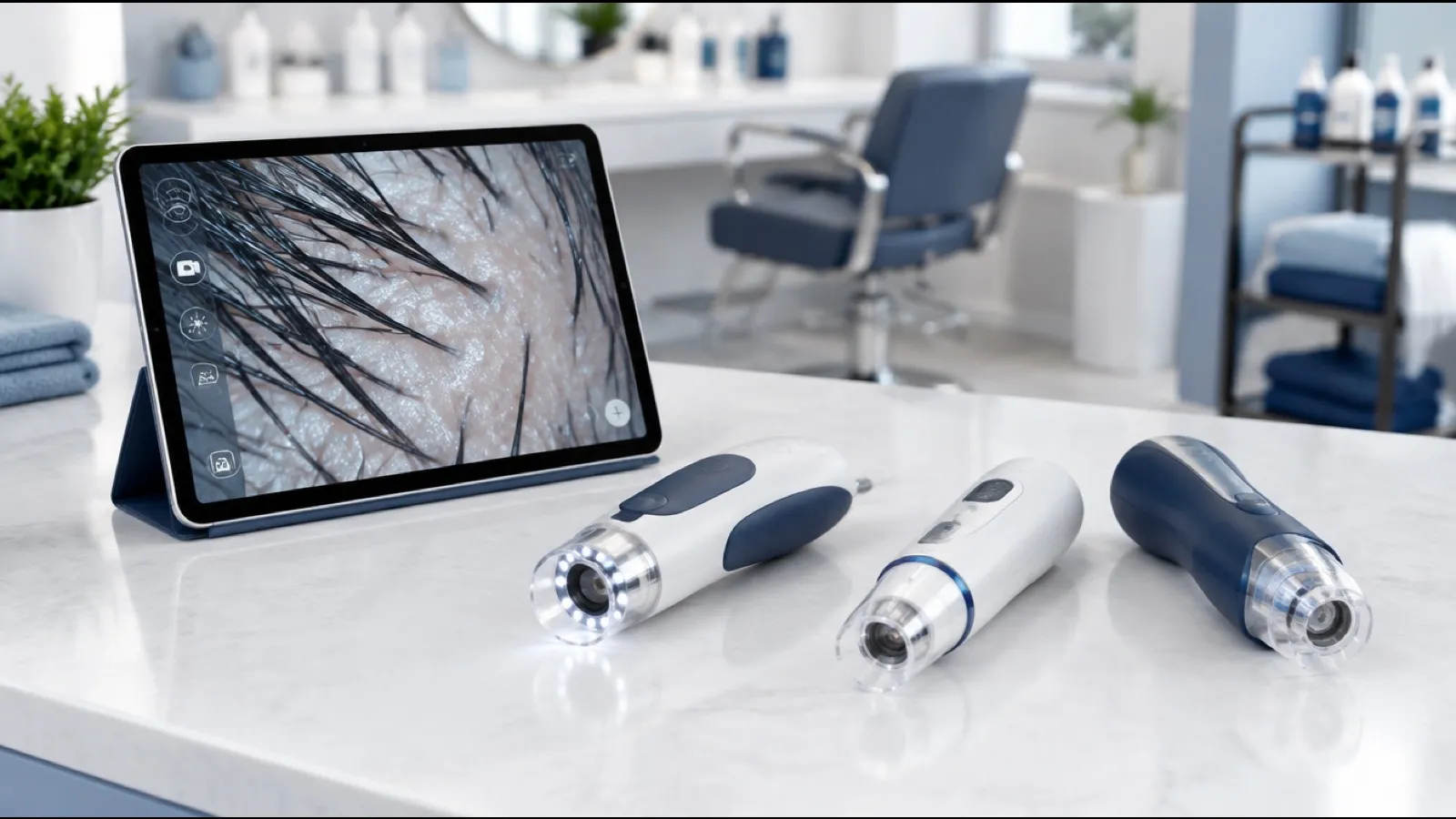
Best Scalp Camera for Salon Use: What to Compare First
The best scalp camera for salon use is the one that gives clear, repeatable images. Compare magnification, lighting, storage, display and tip hygiene.

Hair and Scalp Consultation: Full Guide and Intake Form
A hair and scalp consultation in six stages, with a ready-to-copy intake question list, an observation checklist, summary wording and a follow-up schedule.
How to Talk to a Client About Hair Loss: 6 Salon Scripts
How to talk to a client about hair loss: word-for-word scripts for six salon situations, a say-this-not-that table, privacy rules and referral wording.